Vecteezy logo
Vecteezy logo
Toggle filters
Vectors
Expand vectors navigation
Trending Searches
Top Searches
Backgrounds
Banners
Plants
Flowers
Pattern
Wedding
People
Landscape
Photos
Expand photos navigation
Trending Searches
Top Searches
Nature
Lifestyle
Animals
Food & Drink
Travel
Business
Textures
Cityscapes
Videos
Expand videos navigation
Trending Searches
Top Searches
Family
Timelapses
Animals
Travel
Lifestyle
Aerials
Nature
Backgrounds
Templates
Bundles
More
Expand more navigation
SVGs
Logos
Flowers
Hearts
Arrows
See more SVGs
PNGs
Flower
Frame
Heart
Tree
See more PNGs
PSDs
Logos
Banners
Text Effects
Business Cards
See more PSDs
My Collections
Plans
Plans
Vectors
Trending Searches
Backgrounds
Banners
Plants
Flowers
Pattern
Wedding
People
Landscape
Vector Pages
Homepage
Top Searches
Photos
Trending Searches
Nature
Lifestyle
Animals
Food & Drink
Travel
Business
Textures
Cityscapes
Photo Pages
Homepage
Top Searches
Videos
Trending Searches
Family
Timelapses
Animals
Travel
Lifestyle
Aerials
Nature
Backgrounds
Video Pages
Homepage
Top Searches
Bundles
More
SVGs
PNGs
PSDs
My Collections
Sign Up
Free
Log In
0
Plans
Sign Up
Free
Log In
Photos
Expand filters
All Images
Photos
PNGs
PSDs
SVGs
Templates
Vectors
Videos
Motion Graphics
Search by Image
giant cell arteritis
Search
Search by Image
Explore Other Popular Photo Searches
Recent searches
Reset color
Toggle filters
Photos
Expand filters
All Images
Photos
PNGs
PSDs
SVGs
Templates
Vectors
Videos
Motion Graphics
Search by Image
giant cell arteritis
Search
Search by Image
Explore Other Popular Photo Searches
Recent searches
Reset color
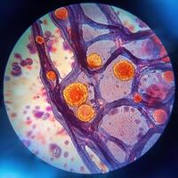
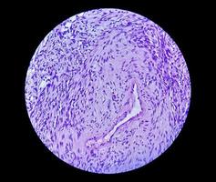
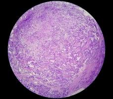
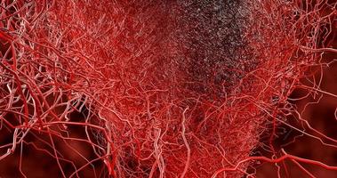
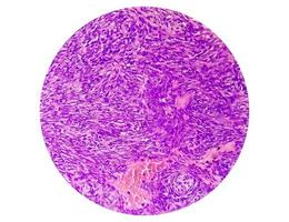
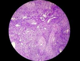
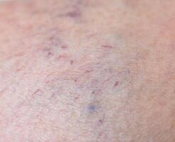
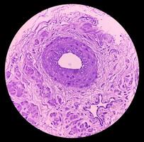
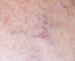
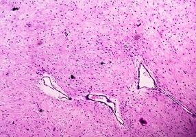
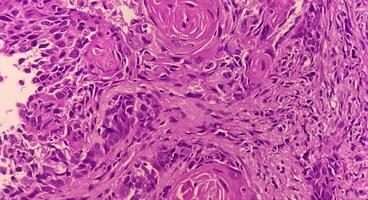
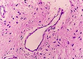
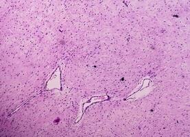
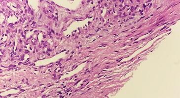
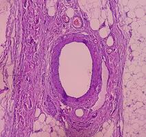
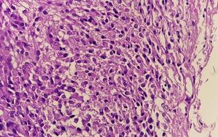
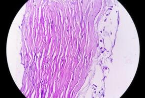
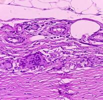
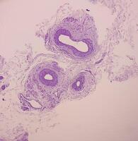
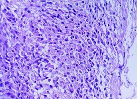
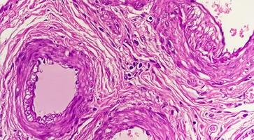
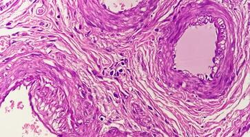
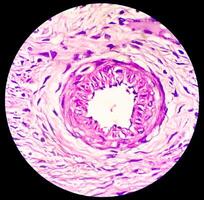
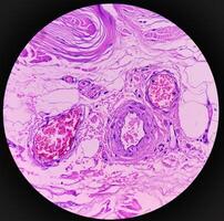
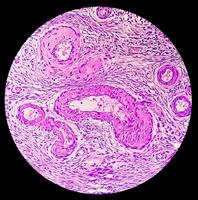
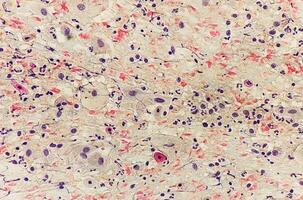
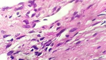
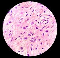
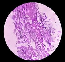
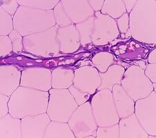
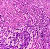
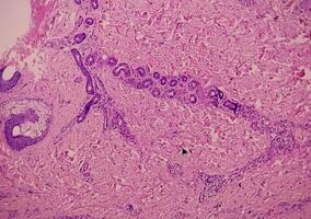
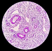
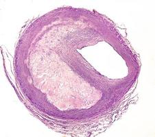
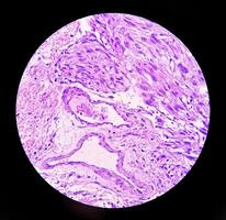
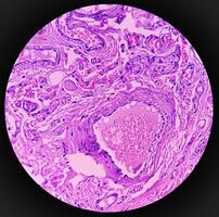
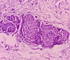
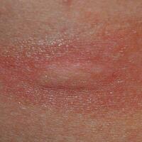
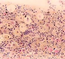
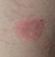
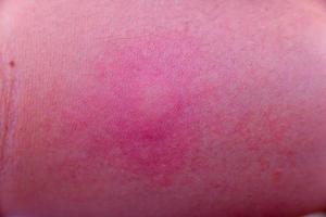
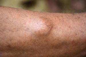
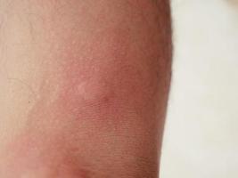
Giant Cell Arteritis Photos & Images
-
205 high resolution, royalty free stock photos and pictures matching
Giant Cell Arteritis
Previous
1
Next
of 3
View More
Vectors
Videos
PNGs
conjunctivitis
coronary artery disease
blood vessel
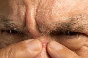
arthritis
eye disease
eye veins
blood clot
skin rashes
epithelial tissue
atherosclerosis
skin cells
sinusitis
cardiovascular system
cardiovascular disease
glaucoma
fungal infection
nasal cavity
osteoarthritis
rheumatoid arthritis
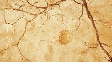
central nervous system
nerve cell
anemia
Previous
Next
Free
Free
cardiovascular disease
glaucoma
fungal infection
nasal cavity
osteoarthritis
rheumatoid arthritis
central nervous system
nerve cell
anemia
varicose veins
tonsillitis
vascular
Free
coronary artery disease
blood vessel
arthritis
eye disease
eye veins
blood clot
skin rashes
epithelial tissue
atherosclerosis
skin cells
sinusitis
cardiovascular system
Free
Free
Free
Free
Free
Free
Free
Free
Free
Free
Click to view uploads for {{user_display_name}}
{{contributor_username}}
{{contributor_resource_count}} Resources
{{follow_button_text}}
Click to view uploads for {{user_display_name}}
{{user_display_name}}
Bookmark icon
Intersect icon
Popular Searches in the US
mothers day
softball
flower background
water
Free Download for Pro Subscribers!
1600 Backgrounds Bundle
View & Download
Available For:
1
Days
11
Hours
36
Mins
18
Secs
Sign Up Free
Already a member?
Log In →
Sign up with Google
Sign up with Facebook
or
Sign Up with Email
Choose a Password
Sign Up Free
Log In to Vecteezy
Login with Google
Login with Facebook
or
Username/Email Address
Password
Log In
Reset Password
Account Email Address
Reset Password
Back to Log In →
Single Sign-on
Log in with your team's identity provider:
Your Vecteezy Team ID
Continue
← Back to Log In